Master Scientific Illustration
In this international study programme you will learn to make accurate visualizations of topics from the clinical, medical and biological domain. You will get the skills to use a wide range of traditional and digital visualization techniques.
It is unique in Europe!
- Location Maastricht
- Duration 2 years
- Degree Master of Arts
- Language English
About the education
One image tells more than a thousand words
It is not surprising that in biomedical sciences traditionally illustrations are created to enable communication between scientist and author, teacher and student, or physician and patient. Art and science come together in scientific illustration.
Best of two top universities
The Master Scientific Illustration is an international study programme in which you will meet students from European countries and beyond. It is a cooperation between Zuyd University of Applied Science and the Faculty of Health, Medicine and Life Sciences at Maastricht University.
Language
This study programme is in English. The lecturers also speak Dutch and German.
Study load
Normally you will study and work at the university for about 20 hours a week. In addition you will have to study at home for about 20 hours a week.
Duration
Full-time for the duration of two academic years from September till July.
Location
Scientific Illustration has it's residence at the attic of a beautiful restored former mansion in the city centre of Maastricht. In addition you will quite often have lectures or workshops at Faculty of Health, Medicine and Life Sciences of Maastricht University.
Lecturers
A small team of dedicated professionals, who all have their own specialism, like scientific and medical illustration, anatomy, software, facial reconstruction etc., will train you. Input from their work experience and their involvement in issues from the professional practice keep the programme up-to-date, educationally justified and closely connected to this professional practice. They all regularly publish imagery, contribute to congresses and take part in the debate about the profession of scientific and medical illustration. External professional experts contribute as guest speakers in lectures, workshops and in the final graduation projects.
-
Rogier Trompert MA
-
Dirk Traufelder MA
-
Andreas Herrler
-
Alex Vent
Tuition fees
The tuition fees for this master differ from the usual institutional tuition fees because this is a non-funded study programme. The fees are based on the actual costs.
Education and Examination Regulations
The education and examination regulations provide all the information you will need about the content of the study programme, the academic support provided, and the way in which examinations are organized.
Study structure
The themes
The study programme consists of three themes:
- Man
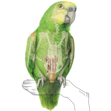
- Animal
- Surgical intervention in man and animal
Each of the themes consists of three to five components and each component involves one or more assignments, which deal with various aspects of scientific illustration and in which various traditional visualization techniques are practised and applied. The assignments are graded in terms of complexity, leading up to the level required for professional practice. Read more about the study programme (pdf).

Training in technical skills
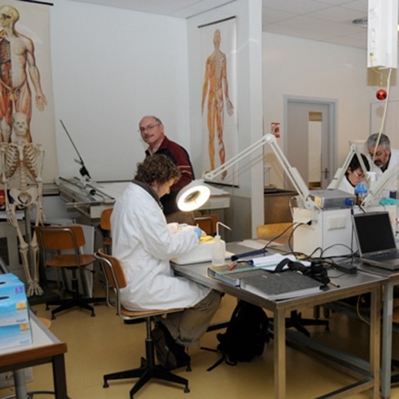
Your training has a strong emphasis on the application of conventional imaging techniques in conjunction with photography, video and computer techniques for accurate two-dimensional display of three-dimensional structures. Additional training in digital three-dimensional reconstruction and modelling is given in workshops. You acquire a broad theoretical basis as well as practical experience in working with medical techniques such as dissection, processing of microscopic and macroscopic serial sections and working with medical imaging techniques such as Computed Tomography (CT) and Magnetic Resonance Imaging (MRI).
Becoming a scientific storyteller
To be able to create a scientifically correct image, you must not only be a good craftsman, but also an outstanding 'storyteller' and communicator. You must be capable of communicating with specialists from different scientific fields, understanding the scientific problem and then be able to convert it into visualization for a specific target audience. Creating images for patient education requires a different approach than creating images for a group of medical specialists. For this reason you will not only be trained in anatomy and medical nomenclature but you’ll also be guided in the field of communication. Furthermore, by means of practical assignments (including illustrating a surgical procedure) you will build up experience in making abstractions and schematisations of the reality to create an image that tells the scientific story in the best possible way.
Graduation
Final examination and graduation
The programme ends with a final examination, consisting of two components:
- A graduation project in which you write, illustrate and design a publication.
- An oral examination.
In addition, you exhibit your work in a graduation exhibition and you compose a portfolio of your representative artworks you produced during the programme. The publication and the portfolio serve as masterpiece that helps you to find work in a professional practice.


 Programme Director